ChemiDoc Imaging Systems
ChemiDoc Imagers offer best-in-class performance with ease of use for visible light (RGB) and far red/near infrared (FR/NIR) fluorescence and chemiluminescence detection and all general gel documentation applications. Stain-free imaging enables immediate visualization of proteins without gel staining and instant verification of protein transfer to blots.
ChemiDoc System Comparison
|
Image
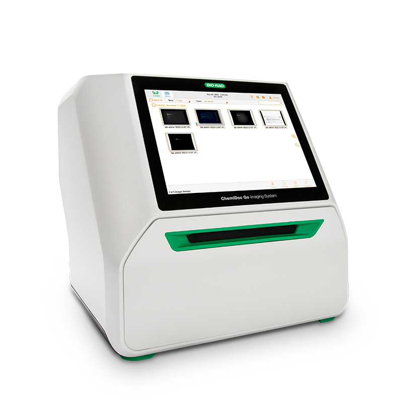
ChemiDoc Go
|
ChemiDoc MP
|
ChemiDoc
|
|
|---|---|---|---|
| Features |
 Modern, compact imaging system with advanced chemiluminescence and StarBright™ Blue fluorescence detection and cloud connectivity. Modern, compact imaging system with advanced chemiluminescence and StarBright™ Blue fluorescence detection and cloud connectivity.
|
High-end imaging system for the best fluorescence and chemiluminescence detection
|
High-sensitivity Chemiluminescence matching X-ray film
|
| Specs | Ordering | Accessories | Specs | Ordering | Accessories | Specs | Ordering | Accessories | |
| Western Blot Sensitivity | Best | Best | Best |
| User Interface | 9.7" graphical touch screen with Image Lab Touch Software | 12" graphical touch screen with Image Lab Touch Software | 12" graphical touch screen with Image Lab Touch Software |
| Multiplex Fluorescence Imaging | Limited, StarBright Blue 520 & 700 compatible | Yes | Upgradable |
| Documentation | Brochure User Guide | Brochure User Guide | Brochure User Guide |
ChemiDoc Recommendations by Application
Blot Detection
| Chemidoc System |
|
|
|
|---|---|---|---|
| Multiplex Fluorescence | StarBright 520 & 700 | Full RGB, far red, near infrared | Upgradable |
| Chemiluminescence | Best | Best | Best |
| SYPRO Ruby Protein Blot Stain* | ✓ | ✓ | ✓ |
| Colorimetric Blots | ✓ | ✓ | ✓ |
| Stain-Free Blots** | ✓ | ✓ | ✓ |
Nucleic Acid Detection
| Chemidoc System |
|
|
|
|---|---|---|---|
| Ethidium Bromide | ✓ | ✓ | ✓ |
| SYBR Stains, GelGreen and GelRed stains, Diamond™ Stain | ✓ | ✓ | ✓ |
| SYBR® Green I or SYBR® Safe Dye | ✓ | ✓ | ✓ |
| Fast Blast™ DNA Stain | ✓ | ✓ | ✓ |
| UView Stain | ✓ | ✓ | ✓ |
Protein Staining (1-D & 2-D Gels)
| Chemidoc System |
|
|
|
|---|---|---|---|
| Stain-Free Gels* | ✓ | ✓ | ✓ |
| Coomassie Brilliant Blue & Coomassie Fluor Orange | ✓ | ✓ | ✓ |
| Silver Stain | ✓ | ✓ | ✓ |
| SYPRO Ruby, Flamingo™, and Oriole™ Fluorescent Gel Stains | ✓ | ✓ | ✓ |
| Pro-Q Stain | ✓ | ✓ | ✓ |
| Cy2, Cy3, Cy5 Label | No | Yes | Upgradable |
* Optimal with low fluorescence PVDF membrane
** Requires Bio-Rad's stain-free gels
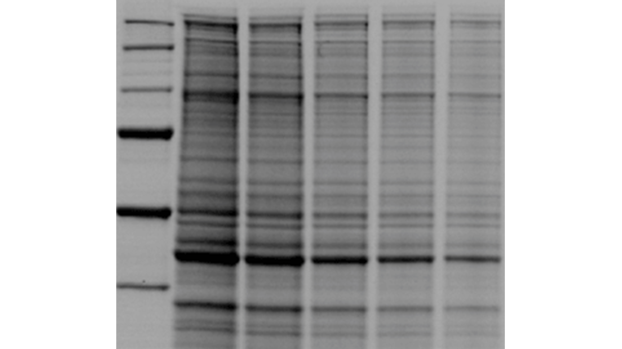
Stain-Free Western Blotting
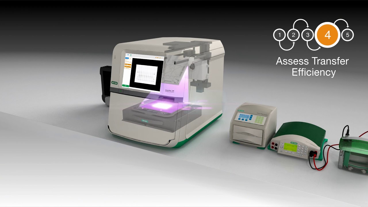
Stain-free western blotting allows you to quickly check electrophoresis and blot transfer quality and obtain truly quantitative western blotting results, updating traditional blotting techniques with innovative tools.
Faster Results. Better Data.
ChemiDoc Go Imaging System: Small Footprint, Big Results
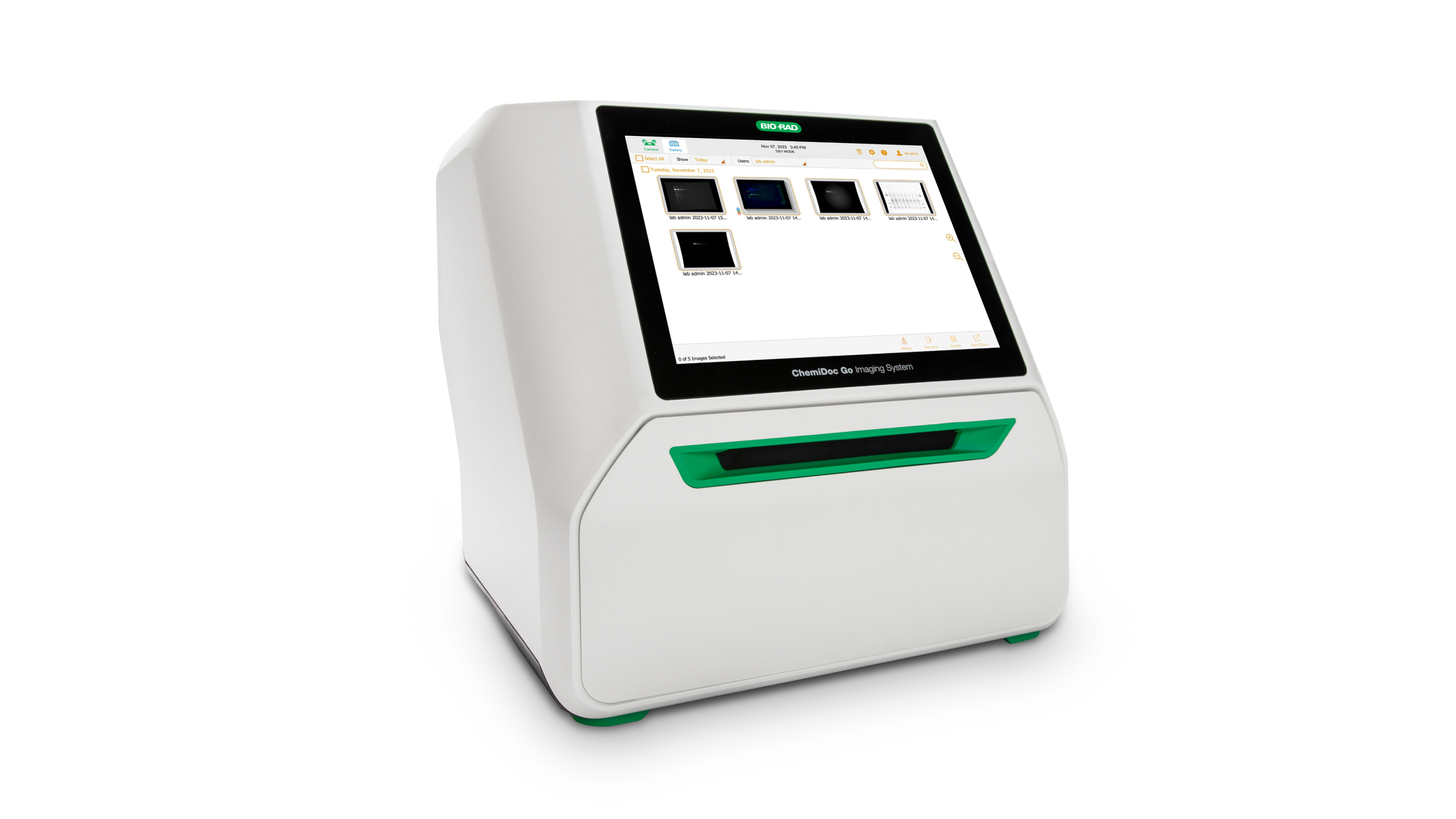
PRODUCT SPOTLIGHTConsidering a GelDoc Go System for your lab?
The ChemiDoc Go Imaging System is capable of imaging chemiluminescence, Stain-Free gels and blots, all common nucleic acid and protein gel stains, as well as StarBright Blue 520 and 700 fluorescent blots. Though compact, the ChemiDoc Go utilizes advanced scientific CMOS digital imaging to capture gel and western blot images with the same sensitivity as larger imaging instruments. It is the first gel and western blot imager capable of connecting to the BR.io cloud for easy image upload, storage, and access.
Benefits of ChemiDoc Imaging Systems
Total Protein Normalization
Learn how to properly design a quantitative western blotting experiment and faithfully compare protein expression levels, without concern for changing or overexpressed HKP’s.
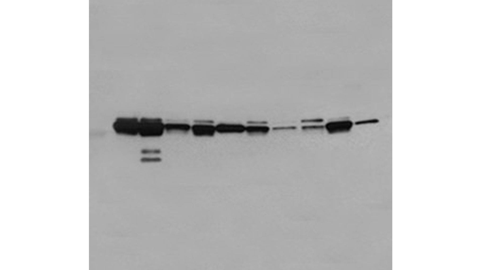
Replaces Film
Save money on consumables and reduce environmental waste without compromising on performance. Get the sensitivity of film and the convenience of digital documentation.
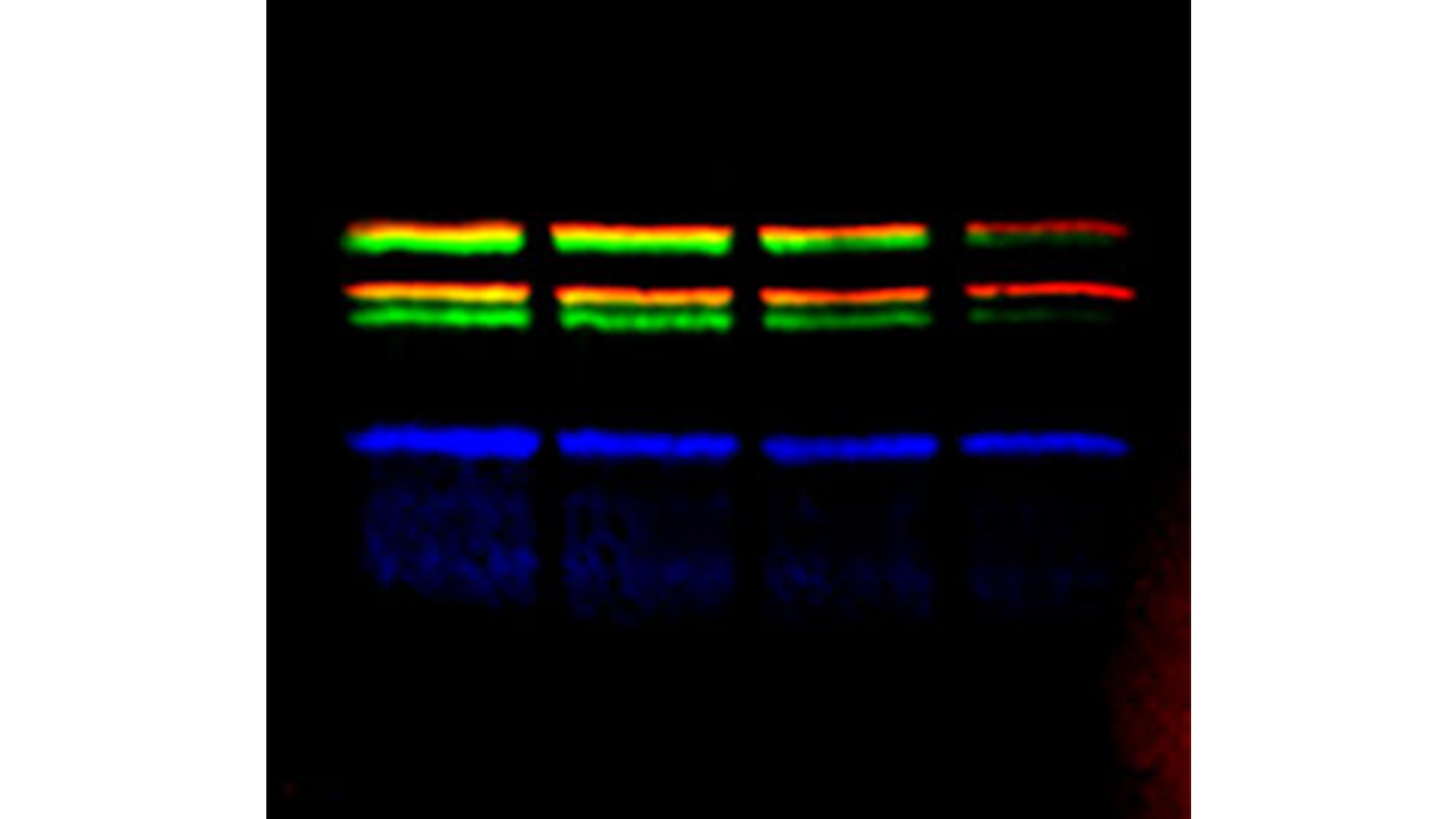
3 Colors
Save sample, avoid the errors commonly associated with stripping, reprobing, and cutting blots and answer more complex biological questions by optimizing your experiments to fluorescence.
Image Lab Software
The Chemidoc System features the intuitive Image Lab Touch Software onboard, with capabilities such as auto image capture, auto analysis, user preferences, and many other features, making gel imaging and analysis incredibly easy.
Image Lab Touch Software Demo
Explore how intuitive it is to acquire fluorescent and chemiluminescent gels and blots on the ChemiDoc systems. This easy-to-use software is perfect for multi-group use and comes installed on the ChemiDoc, ChemiDoc MP, and ChemiDoc Go.

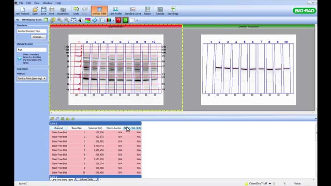
Image Lab Software Introduction
Learn the basics of Image Lab Software for ChemiDoc and Gel Doc Imaging Systems. Find out how to acquire, display, edit and analyze gel and blot images, and then display and export data. This step-by-step webinar is divided into chapters so that you can quickly find the information that you need.
Using Software for Total Protein Normalization on Western Blots
Get an in-depth look at how to normalize your multiplex fluorescence western blots to total protein signal. This video covers manual adjustment of lanes and bands, correcting for blot background, and how to export data for publication or further analysis of your results.

Western Blot Normalization Using Image Lab Software
(PDF 444 KB)
Imaging Fluorescently Stained Gels with Image Lab Software
(PDF 582 KB)
Image Lab Touch Software Resources
Bio-Rad provides instructional videos for Image Lab Touch Software 4.0, covering the primary applications of the ChemiDoc Go Imaging System. Topics include, logging in, Smart Tray Technology, capturing your image, and exporting data.
Resources
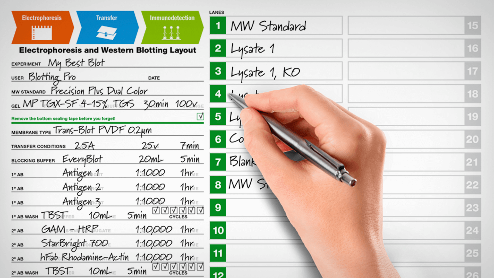
Free 26-Lane Electrophoresis & Western Blotting Layout Post-It Pad
Plan, track, and never miss another step.
Introducing the Western Blot Learning Center
Perfect your western blotting. Learn from the experts.

A Defined Methodology for Reliable Quantification of Western Blot Data
(PDF 602 KB)
Stain-Free Approach for Western Blotting
(PDF 885 KB)
Multiplex Fluorescent Blot Detection Troubleshooting Guide
(PDF 994 KB)
Request a Free Fluorophore Reference Poster
Determine the best fluorophore for maximum sensitivity and high signal-to-noise ratio in your next experiment using this poster.
Applications & Technologies

Western Blotting Doctor
Protocols, video tutorials, and selection guides are available to help you at every step of your electrophoresis experiments.

Horizontal Nucleic Acid Electrophoresis
Learn about gel boxes, running buffers, agarose types, and other factors affecting resolution in DNA gel electrophoresis.

2-D Electrophoresis, Imaging, & Analysis
2-DE is an essential step in protein characterization, purification and profiling, and posttranslational modification studies.

Stain-Free Imaging Technology
Visualize proteins on gels and blots without staining and destaining steps, and use total protein normalization for quantitative western blots.

In-Cell Western Assays
In-Cell Western Assays are high throughput quantitative and qualitative immunofluorescence assays that utilizes Bio-Rad's ChemiDoc MP Imaging System and Image Lab Software.